

Cells Alive Worksheet Answer Key PDF: A Comprehensive Guide
Discover essential resources for mastering cell biology! This guide provides access to Cells Alive worksheets and their corresponding answer keys in PDF format, aiding comprehensive learning․
What are Cells? The Basic Units of Life
Cells, typically microscopic in size, represent the fundamental structural and functional units of all known living organisms․ They are the smallest entities capable of performing life processes, and all living things are composed of one or more cells․ These remarkable structures are not merely building blocks; they are dynamic, self-contained systems capable of growth, reproduction, and responding to their environment․

Some cells, like bacteria, exist as single-celled organisms, carrying out all necessary life functions within that single unit․ Others, such as those found in plants and animals, are part of multicellular organisms, working together in a coordinated manner to form tissues, organs, and ultimately, the entire organism․ The core components of cells – nucleic acids, proteins, carbohydrates, and lipids – are consistent across all life forms, highlighting a shared ancestry and fundamental unity․
Understanding cells is paramount to understanding life itself․ They are the foundation upon which all biological processes are built, and their study is central to fields like medicine, biotechnology, and environmental science․ The study of cells allows us to unravel the complexities of life and develop solutions to pressing global challenges․
Historical Context: From Hooke to Cell Theory
The journey to understanding cells began in 1665 with Robert Hooke, who, using an early microscope, observed compartments in cork and coined the term “cell․” However, Hooke was observing cell walls, not the living contents within․ Further advancements in microscopy throughout the 19th century allowed scientists to observe living cells and their internal structures․
A pivotal moment arrived in 1839 with Matthias Jakob Schleiden’s observation that all plants are composed of cells․ Theodor Schwann extended this finding to animals in 1839, proposing that all living organisms are made of cells․ This formed the cornerstone of the Cell Theory․ Later, Rudolf Virchow added the crucial principle that all cells arise from pre-existing cells, completing the modern formulation of the theory․

This groundbreaking theory revolutionized biology, shifting the focus from organism-level studies to the cellular level․ It provided a unifying framework for understanding the structure, function, and origin of life․ The development of Cell Theory wasn’t a single event, but a gradual process built upon the observations and insights of numerous scientists over time, fundamentally changing our understanding of the biological world․
Prokaryotic vs․ Eukaryotic Cells: Key Differences

Cells fall into two primary categories: prokaryotic and eukaryotic, distinguished by their internal complexity․ Prokaryotic cells, like bacteria and archaea, lack a nucleus and other membrane-bound organelles․ Their genetic material resides in a nucleoid region, but isn’t separated from the rest of the cell․ They are generally smaller and simpler in structure than eukaryotic cells․
Eukaryotic cells, found in plants, animals, fungi, and protists, do possess a nucleus, housing their DNA, and a variety of membrane-bound organelles – mitochondria, endoplasmic reticulum, Golgi apparatus, and more․ These organelles compartmentalize cellular functions, increasing efficiency․ Eukaryotic cells are typically larger and more complex․
A fundamental difference lies in organization; prokaryotes are less organized internally, while eukaryotes exhibit a high degree of internal organization․ This distinction impacts their functions and evolutionary history․ Understanding these differences is crucial for comprehending the diversity of life and the intricacies of cellular processes․ The Cells Alive resources often highlight these distinctions through visual aids and detailed comparisons․
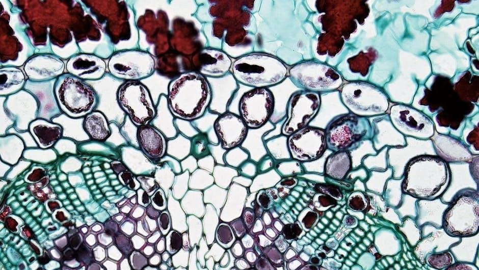
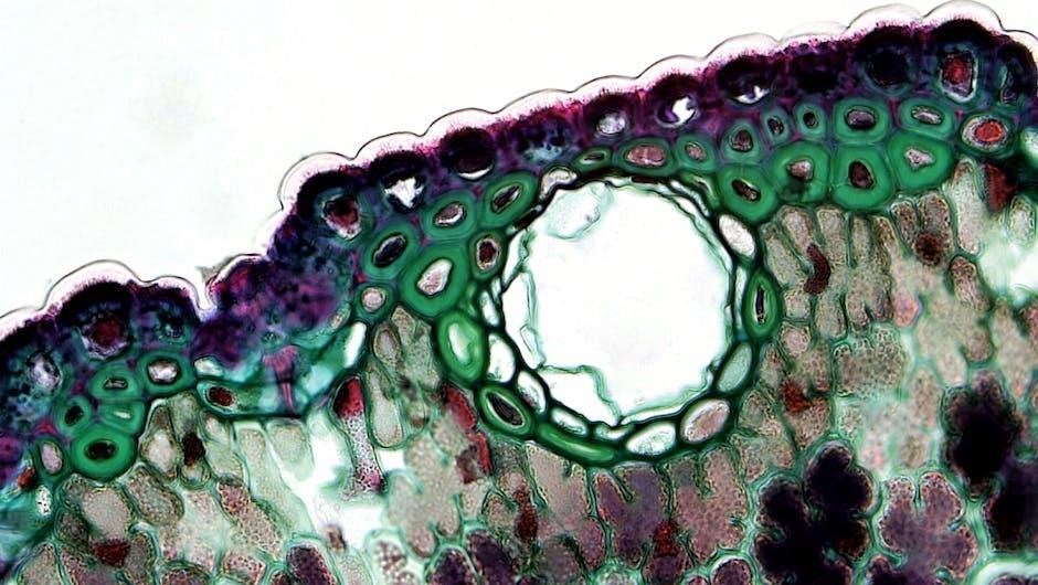
Cell Structures: A Detailed Overview

Cells, the fundamental units of life, aren’t simply homogenous blobs; they’re intricate structures comprised of numerous components working in harmony․ These structures, both internal and external, dictate a cell’s function and contribute to the overall organization of living organisms․ Key components include the cell membrane, providing a protective barrier, and the cytoplasm, the gel-like substance filling the cell․
Within the cytoplasm reside organelles, each with a specialized role․ The nucleus controls cellular activities, while ribosomes synthesize proteins․ The endoplasmic reticulum (ER) facilitates transport and processing, and the Golgi apparatus packages and distributes molecules․ Mitochondria generate energy through cellular respiration, and lysosomes break down waste materials․
Cells Alive worksheets often focus on identifying and understanding the functions of these structures․ Detailed diagrams and interactive models help visualize these microscopic components․ Comprehending these structures is vital for grasping how cells operate and contribute to the complexity of life․ Studying these components provides a foundation for understanding more advanced biological concepts․
The Cell Membrane: Structure and Function
The cell membrane, a vital structure, acts as a selective barrier defining a cell’s boundaries․ Composed primarily of a phospholipid bilayer, it’s a dynamic and fluid mosaic․ Proteins embedded within this bilayer facilitate transport, communication, and structural support․ This membrane isn’t merely a passive enclosure; it actively regulates what enters and exits the cell, maintaining internal homeostasis․
Cells Alive worksheets frequently explore the membrane’s permeability and transport mechanisms – passive diffusion, osmosis, and active transport․ Understanding these processes is crucial for comprehending how cells obtain nutrients, eliminate waste, and respond to their environment․ The membrane’s structure directly influences its function, allowing for selective permeability․
Worksheet answer keys often detail the roles of different membrane proteins, like channel proteins and carrier proteins, in facilitating molecule passage․ Mastering this concept is foundational for understanding cellular physiology and how cells interact with their surroundings․ Visual aids and interactive models enhance comprehension of this complex structure․
The Nucleus: Control Center of the Cell
The nucleus reigns as the cell’s command center, housing the genetic material – DNA – organized into chromosomes․ This double-membrane bound organelle safeguards DNA and orchestrates cellular activities, including growth, metabolism, and reproduction․ Cells Alive worksheets emphasize the nucleus’s role in gene expression and protein synthesis, processes vital for cell function․
Worksheets often depict the nucleus’s key components: the nucleolus (ribosome production), nuclear pores (transport regulation), and chromatin (DNA and protein complex)․ Understanding these structures is crucial for grasping how genetic information is accessed and utilized․ Answer keys provide detailed explanations of these components and their functions․

Cells Alive resources frequently explore the relationship between the nucleus and the cell cycle, highlighting how DNA replication and cell division are meticulously controlled․ Mastering nuclear structure and function is fundamental to understanding heredity and cellular processes․ Interactive diagrams and animations aid in visualizing this complex organelle․
Organelles: Specialized Structures Within Cells
Organelles are the cell’s internal compartments, each performing specific functions essential for life․ Cells Alive worksheets meticulously detail these structures, from energy producers like mitochondria to protein synthesizers like ribosomes․ Understanding organelle roles is key to comprehending cellular processes․
Worksheets often feature diagrams requiring students to identify and label organelles, reinforcing their knowledge of structure and location․ Answer keys provide accurate labeling and detailed descriptions of each organelle’s function․ This includes the endoplasmic reticulum (ER) for transport, the Golgi apparatus for packaging, and lysosomes for waste removal․
Cells Alive resources emphasize the interconnectedness of organelles, demonstrating how they collaborate to maintain cellular homeostasis․ Interactive animations illustrate organelle functions in dynamic detail․ Mastering organelle identification and function is crucial for understanding cell biology, and the provided answer keys ensure accurate learning and comprehension․
Mitochondria: The Powerhouse of the Cell
Mitochondria are often called the “powerhouses” of the cell due to their crucial role in generating energy through cellular respiration․ Cells Alive worksheets dedicate significant attention to these organelles, exploring their unique structure – including the inner and outer membranes and cristae – and function․

Worksheet exercises frequently involve tracing the steps of cellular respiration within the mitochondria, emphasizing the production of ATP, the cell’s primary energy currency․ Answer keys provide detailed explanations of each stage, ensuring students grasp the complex biochemical processes involved․ Diagrams highlight the importance of the mitochondrial membrane for ATP synthesis․
Cells Alive resources often include animations illustrating the electron transport chain and chemiosmosis, further clarifying energy production․ Understanding mitochondrial function is fundamental to cell biology, and the accompanying answer keys offer a reliable guide for accurate learning and assessment of this vital organelle․
Ribosomes: Protein Synthesis Factories
Ribosomes are essential cellular structures responsible for protein synthesis, translating genetic code into functional proteins․ Cells Alive worksheets thoroughly examine these “protein factories,” detailing their composition of ribosomal RNA (rRNA) and proteins․ Students learn about the two ribosomal subunits – large and small – and their roles in the translation process․
Worksheet questions often focus on the steps of translation: initiation, elongation, and termination․ Answer keys provide clear explanations of how mRNA codons are matched with tRNA anticodons, leading to the assembly of amino acid chains․ Diagrams illustrate the ribosome’s movement along the mRNA molecule․
Cells Alive resources frequently differentiate between free ribosomes (synthesizing proteins for use within the cell) and bound ribosomes (attached to the endoplasmic reticulum, producing proteins for secretion or membrane integration)․ The accompanying answer keys ensure students understand the diverse functions and locations of ribosomes within the cell․
Endoplasmic Reticulum (ER): Transport and Processing
The Endoplasmic Reticulum (ER) is a network of membranes crucial for protein and lipid synthesis, as well as transport within the cell․ Cells Alive worksheets delve into the two main types: rough ER (RER) and smooth ER (SER)․ RER, studded with ribosomes, is central to protein folding and modification, while SER lacks ribosomes and focuses on lipid metabolism and detoxification․
Worksheet questions frequently assess understanding of these functional differences․ Answer keys provide detailed explanations of how proteins are processed and transported through the ER lumen, often referencing glycosylation and quality control mechanisms․ Diagrams illustrate the interconnected network of ER tubules and cisternae․
Cells Alive resources emphasize the ER’s role in calcium storage (SER) and membrane production․ The corresponding answer keys clarify how the ER interacts with other organelles, like the Golgi apparatus, to ensure proper cellular function․ Students learn to distinguish between the structural and functional characteristics of RER and SER․
Golgi Apparatus: Packaging and Distribution
The Golgi Apparatus, often visualized in Cells Alive animations, functions as the cell’s processing and packaging center․ Worksheets focus on its structure – flattened, membrane-bound sacs called cisternae – and its role in modifying, sorting, and packaging proteins and lipids for secretion or delivery to other organelles․
Cells Alive worksheet answer keys detail the cis, medial, and trans compartments of the Golgi, explaining how molecules move through these regions undergoing specific modifications like glycosylation․ Questions often assess understanding of vesicle formation and targeted delivery, highlighting the Golgi’s role in exocytosis․
Students learn how the Golgi receives proteins from the ER and further refines them, adding tags that direct them to their final destinations․ Answer keys provide clear explanations of these tagging mechanisms and the importance of the Golgi in maintaining cellular organization․ Diagrams illustrate the flow of molecules through the Golgi apparatus, emphasizing its dynamic nature․
Lysosomes: Cellular Recycling Centers
Lysosomes, depicted vividly in Cells Alive resources, are the cell’s digestive system․ Worksheets emphasize their role in breaking down cellular waste, debris, and foreign materials through enzymatic hydrolysis․ Answer keys detail the acidic environment within lysosomes, crucial for optimal enzyme function․
Cells Alive worksheet questions often explore the origins of lysosomes from the Golgi apparatus and their involvement in autophagy – the process of self-eating, where cells degrade and recycle their own components․ Understanding phagocytosis, where lysosomes digest engulfed particles, is also a key learning objective․
The answer keys clarify how lysosomes contribute to cellular homeostasis by removing damaged organelles and preventing the accumulation of toxic substances․ Diagrams illustrate the process of vesicle fusion between lysosomes and target materials, showcasing the breakdown of macromolecules․ Students learn about lysosomal storage diseases, highlighting the importance of proper lysosomal function for overall health․
Cellular Respiration: Energy Production
Cells Alive worksheets thoroughly cover cellular respiration, the process by which cells convert nutrients into usable energy (ATP)․ Answer keys detail the three main stages: glycolysis, the Krebs cycle, and the electron transport chain, emphasizing their locations within the cell – cytoplasm and mitochondria․

Worksheets frequently pose questions about the role of oxygen in aerobic respiration and the differences between aerobic and anaerobic respiration (fermentation)․ The answer keys explain how glucose is broken down, releasing energy and producing carbon dioxide and water as byproducts․
Cells Alive resources illustrate the importance of mitochondria as the “powerhouse of the cell,” showcasing their inner membrane folds (cristae) which increase surface area for ATP production․ Students learn to trace the flow of electrons through the electron transport chain and understand the role of ATP synthase․ Diagrams and explanations clarify the overall equation for cellular respiration, solidifying comprehension of this vital process․
Cell Division: Mitosis and Meiosis
Cells Alive worksheets provide detailed explorations of cell division, focusing on both mitosis and meiosis․ Answer keys clearly delineate the phases of mitosis – prophase, metaphase, anaphase, and telophase – illustrating chromosome behavior and the formation of daughter cells․ Emphasis is placed on the role of mitosis in growth and repair․
Worksheets contrast mitosis with meiosis, highlighting the key differences in chromosome number and the production of gametes (sex cells)․ The answer keys explain how meiosis results in genetic variation through processes like crossing over and independent assortment․
Cells Alive resources often include diagrams illustrating the stages of meiosis I and meiosis II, clarifying the separation of homologous chromosomes and sister chromatids․ Students learn to compare and contrast the outcomes of mitosis (two identical diploid cells) and meiosis (four genetically unique haploid cells), understanding their respective roles in asexual and sexual reproduction․
Where to Find Cells Alive Worksheets & Answer Keys (PDF)
Cells Alive offers a wealth of resources directly on their official website, www․cellsalive․com․ Here, you’ll find a dedicated section for worksheets covering various cell biology topics, often available for free download in PDF format․ These worksheets are designed to complement their interactive animations and tutorials․
Additionally, many educators compile and share Cells Alive worksheet collections and answer keys on educational platforms like Teachers Pay Teachers․ A quick search will reveal numerous options, some free and others available for purchase․
Furthermore, general science education websites and online learning repositories frequently host Cells Alive-based materials․ Google searches using keywords like “Cells Alive worksheet PDF” or “Cell Biology worksheet answer key” will yield relevant results․ Always verify the source and accuracy of answer keys found outside the official Cells Alive website․
Resources for Further Learning: Cell Biology Websites
Beyond Cells Alive, numerous websites offer comprehensive cell biology resources․ Khan Academy’s Biology section provides free video lessons and practice exercises covering cellular structure and function, ideal for reinforcing worksheet concepts․
The National Human Genome Research Institute (NHGRI) (www․genome․gov) offers detailed information on genes, cells, and heredity, providing a deeper understanding of the molecular basis of life․ BioMan Biology (www․biomanbio․com) features interactive games and tutorials for engaging learning․
For advanced study, explore the resources available from the National Center for Biotechnology Information (NCBI) (www․ncbi․nlm․nih․gov), including scientific publications and databases․ Nature Education (https://www․nature․com/scitable) provides accessible articles on various biological topics․ These sites complement Cells Alive worksheets, fostering a robust understanding of cell biology․